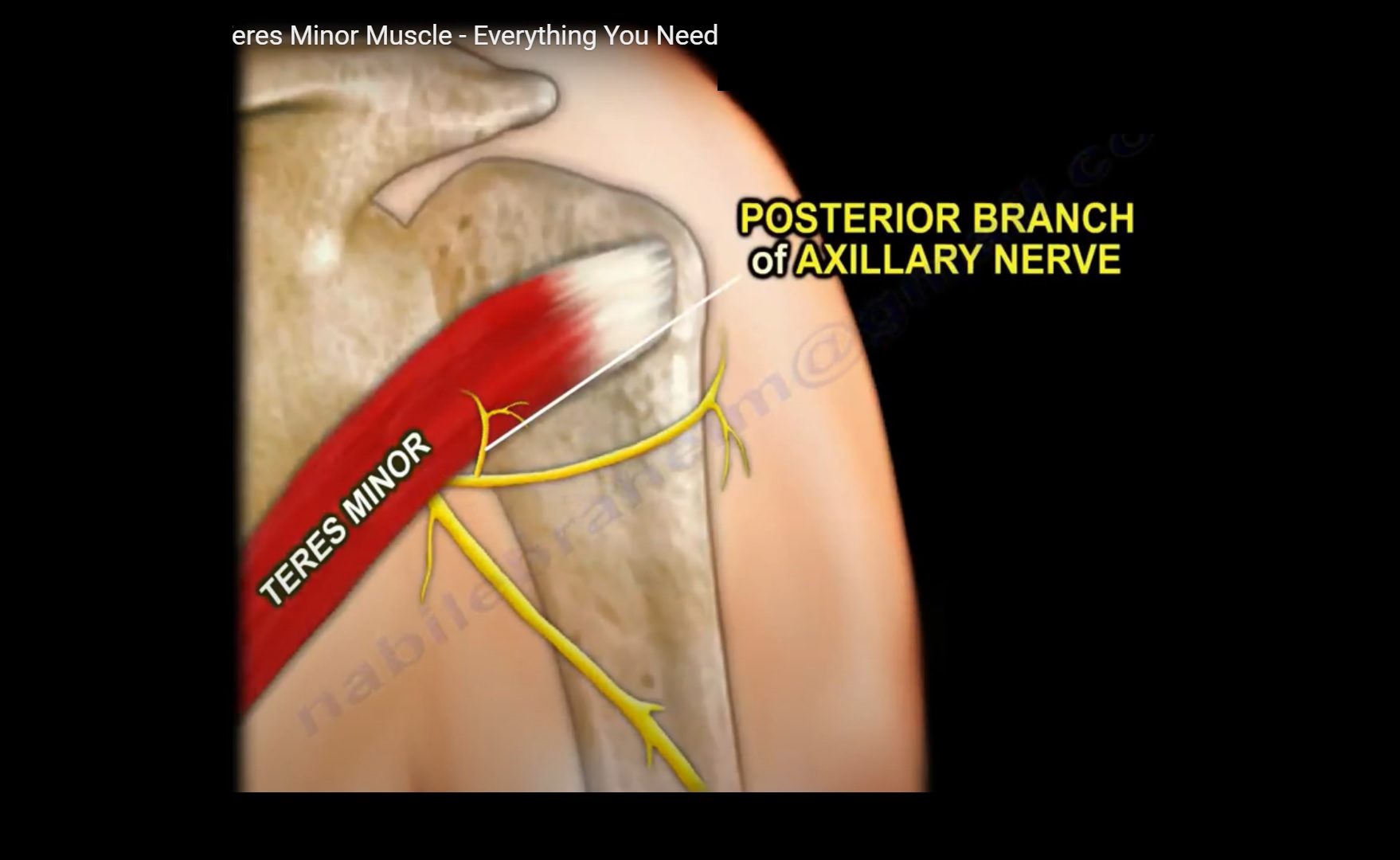


Within the shoulder, the axillary nerve travels posteriorly adjacent to the inferomedial joint capsule before passing through the quadrilateral space beside the posterior circumflex humeral artery. The teres minor muscle is innervated by a branch of the axillary nerve, which originates from the C5 and C6 components of the posterior cord of the brachial plexus. (3C) A fat-suppressed proton density-weighted image below the tendon insertion depicts the broad muscular attachment (arrowheads) of the teres minor along the proximal humeral shaft. (3B) A corresponding sagittal image at the insertion reveals the tendon insertion (arrow) upon the inferior facet of the greater tuberosity (asterisk). (3A) A T2-weighted sagittal image medial to the teres minor insertion demonstrates the central tendon (arrow) within the cephalad aspect of the muscle belly. 4 Muscle fibers of the teres minor also attach broadly to the proximal humerus caudal to the tendon insertion (Figure 3). It has a relatively thin tendon that inserts upon the most inferior of the 3 facets of the greater tuberosity, where it is intimately associated with the posterior capsule. It arises from the dorsal surface of the lateral border of the scapula. The teres minor muscle works in tandem with the infraspinatus as an external rotator of the shoulder, and also assists in dynamic centering of the humeral head. 2, 3 The teres minor muscle is also a site of a characteristic denervation injury at the shoulder, an entity in which our understanding of the process continues to evolve. 1 Alternatively, acute injuries of the teres minor are known to occur following posterior shoulder subluxation or dislocation, in which case concomitant injuries of the infraspinatus may be present. Teres minor tears have commonly been described in the context of large rotator cuff tears in which multiple other cuff tendons tear first.

This is in part due to the fact that tears of the teres minor muscle or tendon are much less common than those of the supraspinatus, the infraspinatus, or the subscapularis.

IntroductionĪlthough rotator cuff tears have been extensively studied and are the focus of a multitude of publications in the scientific literature, the teres minor component of the cuff has received comparatively little attention. Diagnosisįindings compatible with posterior shoulder subluxation with an intramuscular tear of the teres minor, a posterior labral tear, and posterior capsular disruption. (2D) The fat-suppressed T2-weighted coronal image confirms the intramuscular gap (arrow) within the teres minor. An (2C) axial slice inferior to this level shows a tear of the posterior capsule at the humeral attachment (arrow). A posterior labral tear is also apparent (arrowhead). The (2B) fat-suppressed proton density-weighted axial image demonstrates an intact teres minor tendon insertion (arrow) upon the caudal aspect of the greater tuberosity. edition of Gray's Anatomy of the Human Body, published in 1918 – from ).(2A) The T2-weighted sagittal image medial to the rotator cuff insertions reveals an intramuscular gap (arrow) with associated fluid and edema within the teres minor. This definition incorporates text from a public domain edition of Gray's Anatomy (20th U.S. Variations.-It is sometimes inseparable from the Infraspinatus. The tendon of this muscle passes across, and is united with, the posterior part of the capsule of the shoulder-joint. Its fibers run obliquely upward and lateralward the upper ones end in a tendon which is inserted into the lowest of the three impressions on the greater tubercle of the humerus the lowest fibers are inserted directly into the humerus immediately below this impression. The Teres minor is a narrow, elongated muscle, which arises from the dorsal surface of the axillary border of the scapula for the upper two-thirds of its extent, and from two aponeurotic laminae, one of which separates it from the Infraspinatus, the other from the Teres major. Insertion: Inferior facet of greater tubercle of the humerusĪrtery: Posterior circumflex humeral artery and the circumflex scapular arteryĪction: Laterally rotates and adducts the armĪntagonist: Subscapularis, pectoralis major, and latissimus dorsi


 0 kommentar(er)
